Efficient scan patterns for scanning laser microscopy systems
Unmet Need
Scanning imaging systems, such as confocal scanning laser ophthalmoscopy (cSLO), hold great promise in the clinic to diagnose a variety of retinal disease like diabetic retinopathy. These systems are particularly promising for applications in telemedicine and are advantageous over traditional fundus photography, which often do not produce high quality images or are challenging to use. Most existing scanning imaging systems use raster scanning which simplifies the image processing step. However, raster scanning leads to low scan efficiency and poor thermal efficiency (e.g., generating excess waste heat from mechanical scanners), limiting the overall clinical utility of these systems. There is a need for new scanning methods that can match or improve the imaging quality of existing systems, while decreasing the acquisition time and costs to enable a variety of telemedicine applications.
Technology
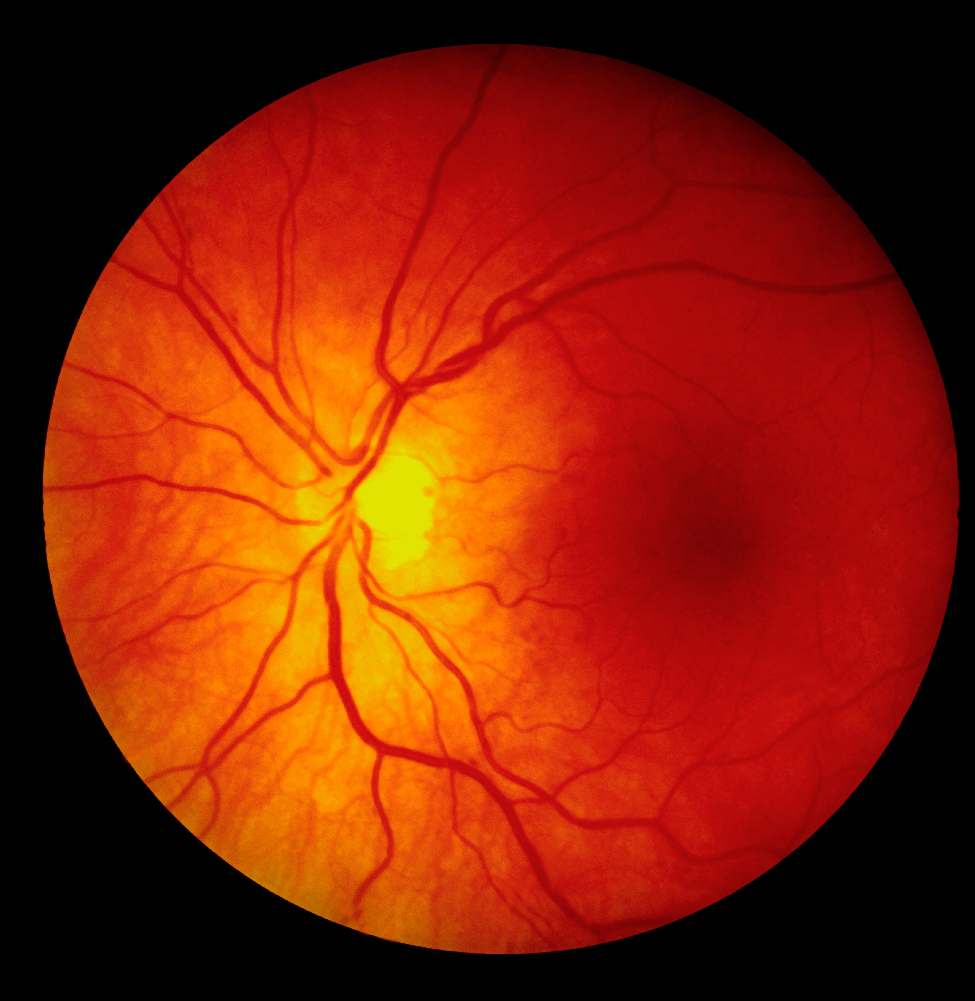
Duke inventors have developed a new hybrid spiral scan pattern that is coupled with image processing techniques to produce high quality images using cSLO. This is intended to be used by ophthalmologists to diagnose a variety of retinal diseases, such as diabetic retinopathy. In addition, the methods could be applied for optical coherence tomography (OCT), laser scanning confocal microscopy (LSCM) imaging systems. Specifically, the imaging system is composed of a green laser diode and NIR superluminescent diode, operating at 520 and 785nm, respectively. The light is collimated, scanned by galvanometers, and relayed into the patient's pupil using off-the-shelf optics and custom optomechanics. The galvanometers use a hybrid spiral scan pattern which includes regions of constant linear velocity spirals and regions of constant angular velocity spirals. Image reconstruction is carried out by an interpolation of collected data to a raster format such that an image can be produced that is suitable for display. The feasibility of this system has been demonstrated by imaging a healthy human volunteer.
Other Applications
This technology could also be applied to other scanning technologies that utilize energy propagating in the form of waves, including all portions of the electromagnetic spectrum (e.g., radio frequency, far-infrared, near-infrared, visible, ultraviolet, and X-ray), and acoustic energy).
Advantages
- The scan method offers performance advantages over traditional raster imaging systems in terms of acquisition speed and costs
- The optical design achieves diffraction-limited performance over a 50° FOV at a working distance of 16mm
- GPU-based algorithms produce contrast-enhanced images in real time
- Light collected from the patient eye is detected on a single photomultiplier tube with no filtering
- Patient safety is ensured by limiting incident power on the subject eye