Improving visualization during ultrasound-guided needle insertions
Value Proposition
Insertion of needles into blood vessels is a common and crucial intervention for insertion of venous and arterial catheters, as well as blood sampling. Similarly, insertion of needles into tumors and other tissues can provide crucial diagnostic information to guide treatment. Use of real-time ultrasound greatly improves the accuracy of these tasks by allowing visualization of the needle entering the target structure, as well as the relationship to surrounding structures. Keeping the needle within the plane of the ultrasound beam is critical for visualization of the needle path, but is highly challenging to do so. Needle guides, consisting of clip-on pieces of plastic that help guide the angle and trajectory of the needle, exist. However, these guides cause the needle to traverse the tissue at an angle, which can encourage skewing of the needle vector and renders it nonvisible. There is a need for a novel ultrasound-guided surgical probe to correct this aberration.
Technology
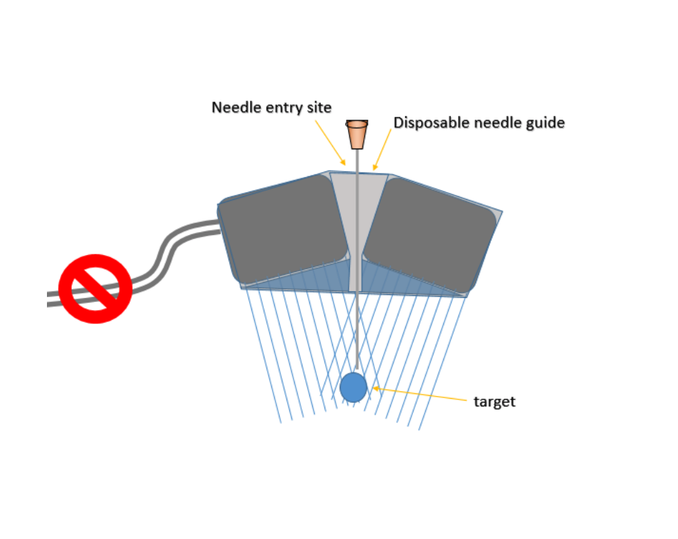
The current technology provides an interventional ultrasound probe that allows for the guidance of a needle within a subject while keeping the needle within the ultrasound beams so that it can be visualized by a caregiver. The design centers on two separate transducers aligned at an angle towards each other. A space in the center is provided for a disposable needle guide. The transducers generate images of the target area in overlapping planes that are then merged in real-time using image processing software. Another embodiment of the technology has a single row of elements set along a concave angle, with central elements removed for room for the needle guide, with generation of a single image.
Advantages
- Reduces complications associated with and improves the speed of ultrasound guided procedures
- Enhanced visualization of needles by virtue of overlapping ultrasound beams, thereby optimizing ultrasound-guidance
- Allows longer area for needles guidance, which will minimize needle skew out of the ultrasound plane